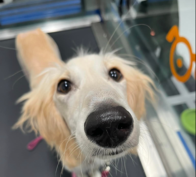
Georgie is a 6-month-old, female, Saluki who presented with a history of poor appetite and soft faeces. On abdominal ultrasonography it was incidentally noted that pleural effusion was present.
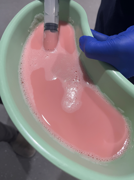
Thoracocentesis revealed opaque fluid grossly consistent with chyle which was later confirmed on fluid analysis and cytology.
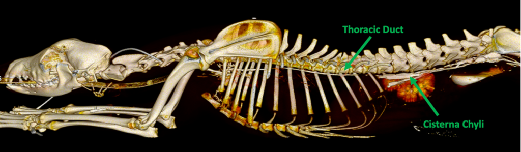
She was also noted to have left thoracic limb oedema which had been a long-standing abnormality. CT lymphangiography was performed which documented the course of the cranial abdominal lymphatics and the thoracic duct as well and a likely congenital malformation of the lymphatics of the left thoracic limb and lateral thoracic wall.
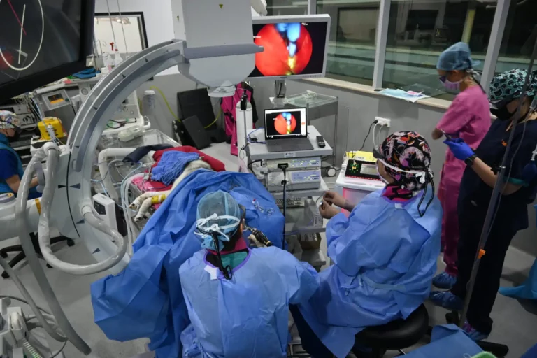
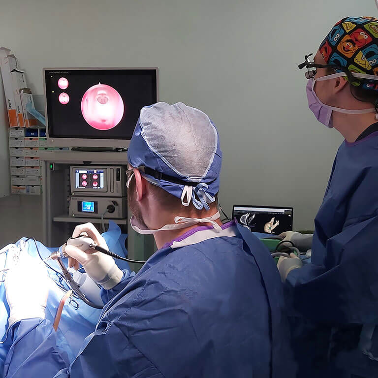
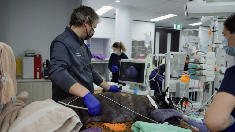
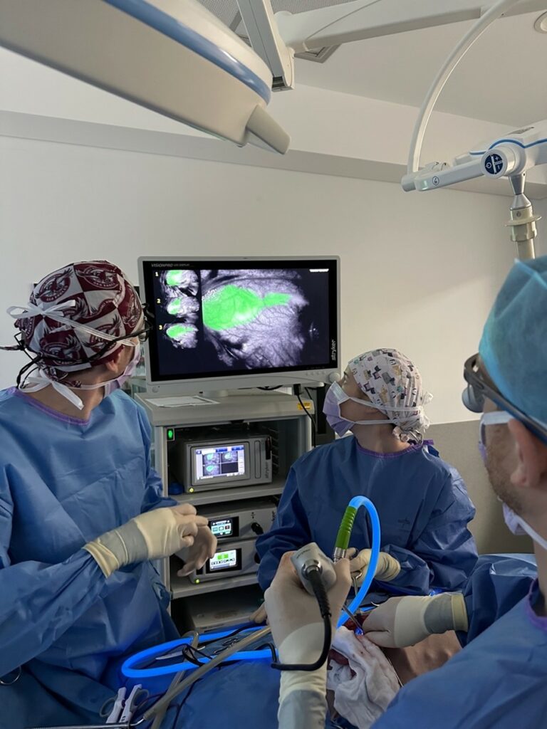
She was taken to surgery and laparoscope assisted near infrared imaging of the cisterna chyli and thoracic duct was performed with indocyanine green dye to visualize the complex anatomy of her lymphatic system. The thoracic duct was then ligated ensuring all tributaries were occluded. Her cisterna chyli was then ablated. This process was greatly aided with the use if of NIRF imaging and ICG and also allowed for a smaller surgical approach.
Georgie’s post-operative recovery was rapid and the volume and character of the pleural effusion improved over two weeks following surgery with resolution of chylothorax. She is now back to her normal self.

If you have a patient you think might benefit from this technology or want to learn more please feel free to contact me at SASH.